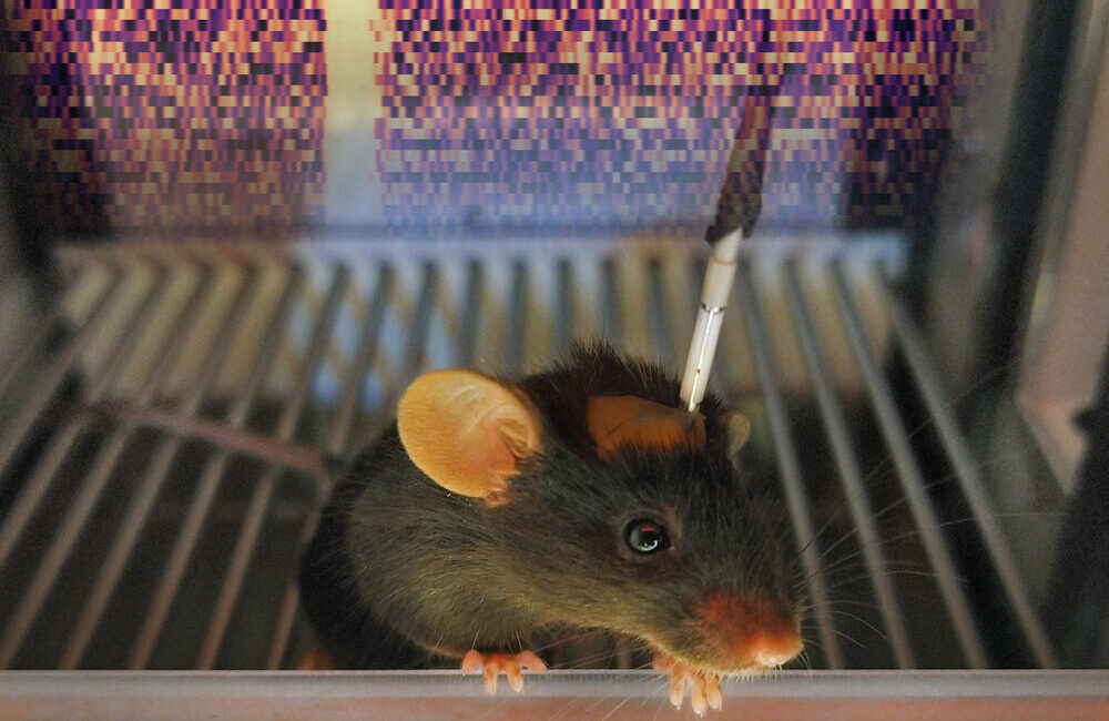
How is neuromodulatory information generated and disseminated throughout the brain? How do individual variations in this circuitry create different moods, personalities, or reward-learning strategies?

 
Lights, camera, action! Peeking into the brains of mice to understand reward learning

Lights, camera, action! Peeking into the brains of mice to understand reward learning


Variability in motivated behavior and the dopamine system across individuals

The midbrain dopamine system is highly plastic. The plasticity of this system is most often explored in the context of drug addiction, yet genetic differences and natural environmental changes also profoundly influence it. We know, for example, that conditions such as environmental enrichment and physical activity influence the number of dopamine neurons in the brain, that adolescent and adult mice differ in their risk-taking behavior, particularly in response to social cues, and that motherhood changes females’ responses to pup calls, likely in a dopamine-dependent manner. The Lerner Lab takes advantage of these important biological sources of behavioral variability to precisely dissect links between midbrain dopamine system structure and function.

Computational Principles for Decision-MakinG

Below is a talk by Dr. Lerner at the 2024 Kavli Frontiers of Science Korean-American Symposium, which provides an overview of the field of decision-making and the computational principles by which we can begin to integrate findings about decision-making from across neuroscientific subdisciplines. She then focuses on examples from the Lerner laboratory, using rodents as a model system to dissect behavioral strategies underlying decisions to seek rewards under the threat of punishment.


Generation of Dopamine Signals

Dopamine signals are generated through a combination of afferent input signaling and intrinsic processing by the dopamine neurons themselves. To examine how dopamine neurons integrate information from afferents, we use the rabies-mediated tracing technique TRIO in combination with whole-brain CLARITY and CLARITY-Optimized Light-Sheet Microscopy (COLM) imaging, as seen in the videos below.

Whole-brain lableing of inputs to DLS-projecting dopamine neurons visualized using CLARITY and CLARITY-Optimized Light-Sheet Microscopy (COLM)

High-Resolution (25x) detail of labeled inputs to DLS-projecting dopamine neurons using COLM

We can then manipulate activity in these anatomically identified inputs using optogenetics and examine the effects of perturbing these inputs on both dopamine neuron firing patterns and behavioral output. Additional electrophysiological investigations can determine the extent to which differences in the intrinsic properties of dopamine neurons contribute to alternations in the generation of dopamine signals.

The information gained from our studies on the sources and strengths of inputs to different classes of dopamine neurons, as well as their intrinsic properties, will inform thinking about how the brain continually adjusts dopaminergic feedback signals in response to changing conditions or internal cues.

TRIO labeling (green) can be used to query whole-brain inputs to specific subsets of midbrain dopamine neurons. Here, we observe that inputs to dopamine neurons from the striatum arise from dopamine D1 receptor-expressing cells (red).

TRIO labeling (green) can be used to query whole-brain inputs to specific subsets of midbrain dopamine neurons. Here, we observe that inputs to dopamine neurons from the striatum arise from dopamine D1 receptor-expressing cells (red).

Whole-cell patch clamp electrophysiology can be used to measure synaptic inputs to dopamine neurons as well as their intrinsic properties. Shown is a patched dopamine neuron visualized using DIC microscopy (left) and a synaptic current measured from…

Whole-cell patch clamp electrophysiology can be used to measure synaptic inputs to dopamine neurons as well as their intrinsic properties. Shown is a patched dopamine neuron visualized using DIC microscopy (left) and a synaptic current measured from a dopamine neuron when striatal inputs to that neuron were stimulated using channelrhodopsin at the time indicated by the blue bar (right).

Dissemination of Dopamine Signals

Are dopamine neurons readily divisible into neat subsets targeting distinct functional output nuclei, or do they tile their target regions following some other organizational rule? Do patterns of dopaminergic innervation in the cortex, amygdala or striatum differ between the behaviorally diverse groups of mice described above or evolve in response to changes such as slow dopamine neuron degeneration in Parkinson’s disease? The Lerner Lab seeks to answer these questions using an array of approaches, including anatomical methods, electrophysiology, multi-fiber photometry, imaging, and optogenetics.

CAV-cre is a viral method that allows us to identify dopamine neurons by projection target. Here, CAV-cre was injected into the dorsomedial striatum (DMS) of a tdTomato reporter mouse. Therefore, DMS-projecting neurons are labeled in red. Dopam…

CAV-cre is a viral method that allows us to identify dopamine neurons by projection target. Here, CAV-cre was injected into the dorsomedial striatum (DMS) of a tdTomato reporter mouse. Therefore, DMS-projecting neurons are labeled in red. Dopamine neurons (identified by immunostaining for TH) are shown in blue.

Red retrobeads are another method that allows us to identify dopamine neurons (here shown in green in a TH-GFP transgenic mouse) based on their output target. In this case, red retrobeads were injected in the dorsomedial striatum (DMS), labeling DMS…

Red retrobeads are another method that allows us to identify dopamine neurons (here shown in green in a TH-GFP transgenic mouse) based on their output target. In this case, red retrobeads were injected in the dorsomedial striatum (DMS), labeling DMS-projecting dopamine neurons in the midbrain. The patterns of labeled cells are similar to those observed using CAV-cre (above).

An example of fiber photometry signals observed in SNc dopamine neurons axons in the dorsomedial striatum (DMS). Mice made nosepoke responses in a behavioral chamber to receive sugar pellets. The signals recorded from DMS dopamine axons in response to rewarded nosepokes (that triggered the delivery of a sucrose pellet) and unrewarded nosepokes (which did not result in a sugar pellet) could be used to predicted the number of shocks mice would tolerate when presented with a risk of footshock punishment for nosepoking. These data demonstrate that individual differences in punishment-resistant reward-seeking can be explained, at least in part, by differences in dopamine circuit function between individual subjects.